

Advanced Dental Imaging
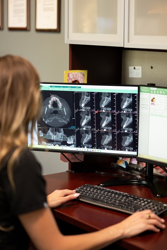
What is CBCT Imaging?
Some procedures require more information than a traditional dental X-ray can provide.
Cone Beam Computed Tomography, or CBCT, creates detailed three-dimensional images of your teeth, jaws, and surrounding anatomy, allowing your surgeon to see structures that cannot always be fully evaluated on a standard 2D X-ray.
Better imaging. More information. Thoughtful surgical planning.
What Does a CBCT Show?
A CBCT scan can provide detailed information about:
- Teeth and tooth roots
- Jawbone anatomy
- Impacted teeth
- Important nerves
- Sinuses
- Available bone for dental implants
- Cysts and other areas of concern
- Complex dental and skeletal anatomy
When Is CBCT Used?
3D imaging may be recommended when planning or evaluating:
- Wisdom tooth removal
- Dental implants
- Bone grafting
- Sinus lifts
- Impacted teeth
- Oral pathology
- Other complex surgical procedures
What to Expect
A CBCT scan is quick, comfortable, and non-invasive.
You’ll stand or sit still while the scanner moves around your head to capture the images. The scan itself typically takes less than a minute.
Your surgeon can then review the 3D images with you and use them to help plan your care.
Why 3D Imaging Matters
Every patient’s anatomy is different.
CBCT imaging allows us to evaluate important structures in three dimensions and plan your procedure based on your individual anatomy rather than relying solely on a traditional two-dimensional image.
Advanced Technology. Personalized Care.
When 3D imaging is appropriate, it gives us another tool to carefully evaluate your anatomy, explain your treatment, and plan your procedure.